Floaters are shadows of opacities in the vitreous.
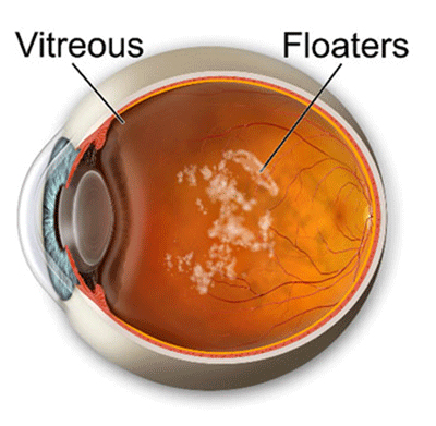
Patients complain of small specks, dots or strands that can be seen more easily against a bright background, such as a diffusely illuminated wall or sky. They are sometimes mistaken for flies, as they often dart around as the eye is moved.
Floaters, if present for a long time or increase in number very gradually, are harmless. They are annoying, but do not cause any problems. If a new floater comes on abruptly, however, or if there is a sudden increase in the number of floaters, then one must be examined promptly to rule out a retinal tear or detachment.
Floaters are commonly due to condensations of the vitreous collagen and are formed as the vitreous degenerates and liquefies as we age. They may also be due to blood (where they typically present as multiple small floaters) usually from a retinal tear, or from glial tissue torn from an area adjacent to the optic nerve head (where they typically present as a large single or a few large floaters) from a posterior vitreous detachment.
Cause: As we age, the consistency of the gel molecules of the vitreous changes as they release their water portion and lacunae (pockets of liquefied vitreous) are formed. The collagen or dense gel-like filaments aggregate to form larger fibrils.
This process causes a collapse of the vitreous gel structure and is known as vitreous degeneration and syneresis. The collagen fibrils “float” within the liquid pockets, giving the patient the sensation of floaters. When the vitreous pulls on the retina, where it is attached, the light-sensing cells are mechanically stimulated, causing a “flash” of light. If it pulls on a blood vessel, it may cause it to leak blood cells which may cause reduced vision and additional floaters. If this occurs, the risk of a retinal tear or detachment is increased.
Vitreous degeneration and syneresis can cause a complete collapse of the vitreous. A posterior vitreous detachment is the term used when this occurs. It happens when enough of the lacunae (pockets of liquid vitreous) accumulate to cause a collapse of the vitreous framework and a separation of the vitreous from the retina. Posterior vitreous detachments occur in less than 10% of people under 50 years of age, but in more than 60% over the age of 70. It is more common in people who are nearsighted, who have had an eye injury, eye inflammation or undergone eye surgery.
Treatment: Over time, you will become less aware of these floaters as the brain learns to ignore these retinal images. Therefore, while some floaters may remain, many will fade over time and become less bothersome. You are advised not to ignore symptoms of sudden increase in the number of floaters or flashes of light, especially if accompanied by subjective reduction in vision (cloud or curtain), as these symptoms can signify an acute posterior vitreous detachment. More seriously, there could be an associated tear or retinal detachment.
A prompt eye examination is needed, where dilating drops will be instilled to open the pupil to obtain a complete view of the retina and rule out any retinal involvement. With no retinal involvement, one must still be aware of the three main signs & symptoms of a retinal tear or detachment:
1- Flashing lights (indicating new traction or pulling on the retina)
2- Increase in floaters or new floaters (indicating new opacities and new activity)
3- Change in vision or a curtain in vision (indicating a possible tear or detachment).
If any of these occur, you are advised to see your eye care professional promptly. You may also be advised to set up a follow-up visit, regardless of signs or symptoms, if your eye doctor feels you are at risk for potential changes.
Back
700 University Avenue, Toronto, Ontario M5G 1Z5
Phone: (416) 977-8194
Monday to Thursday: 8:30am to 5:30pm
Friday: 8:30am to 4:00pm
Saturday & Sunday: Closed